
Direct pathway neurons in mouse dorsolateral striatum in vivo receive stronger synaptic input than indirect pathway neurons
Abstract
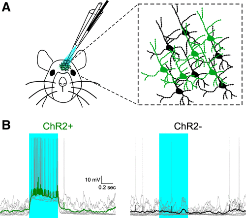
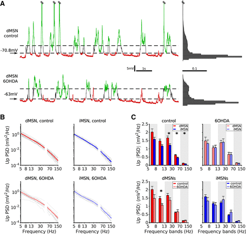
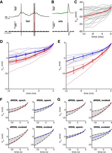
Striatal projection neurons, the medium spiny neurons (MSNs), play a crucial role in various motor and cognitive functions. MSNs express either D1- or D2-type dopamine receptors and initiate the direct-pathway (dMSNs) or indirect pathways (iMSNs) of the basal ganglia, respectively. dMSNs have been shown to receive more inhibition than iMSNs from intrastriatal sources. Based on these findings, computational modeling of the striatal network has predicted that under healthy conditions dMSNs should receive more total input than iMSNs. To test this prediction, we analyzed in vivo whole cell recordings from dMSNs and iMSNs in healthy and dopamine-depleted (6OHDA) anaesthetized mice. By comparing their membrane potential fluctuations, we found that dMSNs exhibited considerably larger membrane potential fluctuations over a wide frequency range. Furthermore, by comparing the spike-triggered average membrane potentials, we found that dMSNs depolarized toward the spike threshold significantly faster than iMSNs did. Together, these findings (in particular the STA analysis) corroborate the theoretical prediction that direct-pathway MSNs receive stronger total input than indirect-pathway neurons. Finally, we found that dopamine-depleted mice exhibited no difference between the membrane potential fluctuations of dMSNs and iMSNs. These data provide new insights into the question of how the lack of dopamine may lead to behavioral deficits associated with Parkinson’s disease.
NEW & NOTEWORTHY The direct and indirect pathways of the basal ganglia originate from the D1- and D2-type dopamine receptor expressing medium spiny neurons (dMSNs and iMSNs). Theoretical results have predicted that dMSNs should receive stronger synaptic input than iMSNs. Using in vivo intracellular membrane potential data, we provide evidence that dMSNs indeed receive stronger input than iMSNs, as has been predicted by the computational model.
